Environmental scanning electron microscope
Object No. 2010/26/1
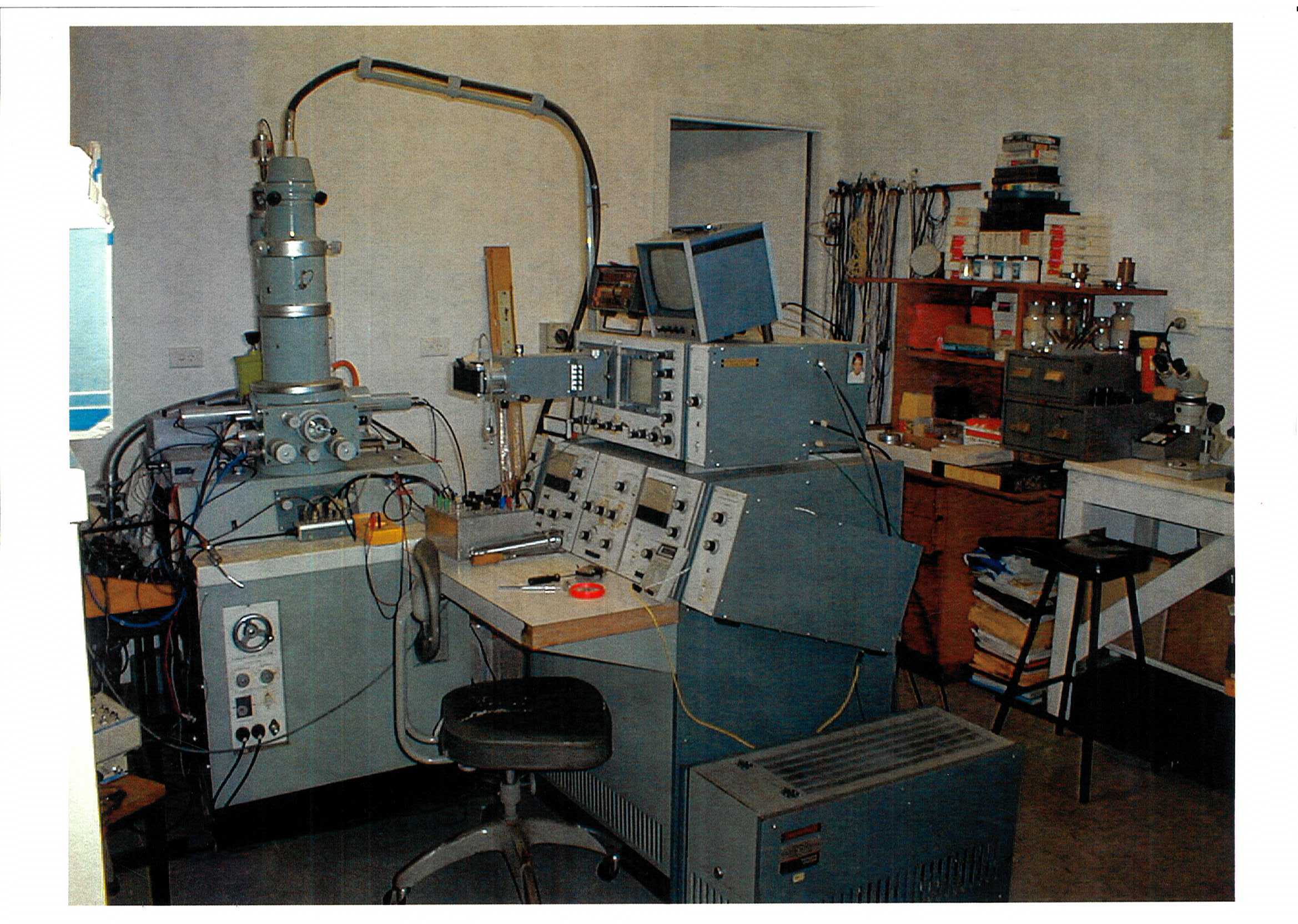
This electron microscope is the first environmental scanning electron microscope (ESEM) in the world. Environmental Scanning Electron Microscopy is an Australian innovation and was invented in Australia on this machine. ESEM units enable the study of objects at high magnification (up to greater than X 100,000) in their natural state or under their natural environmental conditions without destroying the object, and without an applied coating such as sputtered gold, which can destroy sensitive objects. Samples can be studied over a temperature range from -200 C to 1000 C as appropriate. There are now thousands of ESEMs in use world-wide. The significant advancement in electron microscopy that the ESEM provided is that it can scan a sample without it having to be in a vacuum environment. A scanning electron microscope (SEM), scans a sample that is held in a high vacuum environment and coated in a conductive material, usually gold. The vacuum is required so that the scanning beam is not scattered by collisions with gas molecules. The ESEM solved this issue by using a significantly higher pressure sample chamber. The specimen chamber is isolated by valves or pressure limiting apertures and a large diameter bypass tube from the rest of the vacuum system. In addition, new detectors were invented and developed that could operate in a gaseous environment. Water vapour is commonly used as the imaging gas, and thus much less damage is caused to the sample. ESEMs have been used to examine dynamic physicochemical properties during the swelling and dissolution of drugs and the mechanism of drug release; to study the effects of wetting and drying on wool, cotton and synthetic fibres; to follow the reaction of cement with water; to observe fresh and wet tissue such as muscle cells and connective fibres; to examine live plant seedlings, pollen, grains in situ, ants, fleas, dust mites; to investigate the degradation processes caused by environmental pollutants on paintings, sculptures and other artworks; and in investigations for soil science, polymer structure, petroleum geology, archaeology, palaeontology, ceramics, food technology, microelectronics, dentistry, microbiology, and paper coating technology. http://www.danilatos.com/ Damian McDonald February 2010
Loading...
Summary
Object Statement
Environmental scanning electron microscope, prototype, steel / aluminium / plastic / rubber / glass, modified JEOL JSM2 (1968), made by Dr Gerry Danilatos, Australia, 1978 - 2003
Physical Description
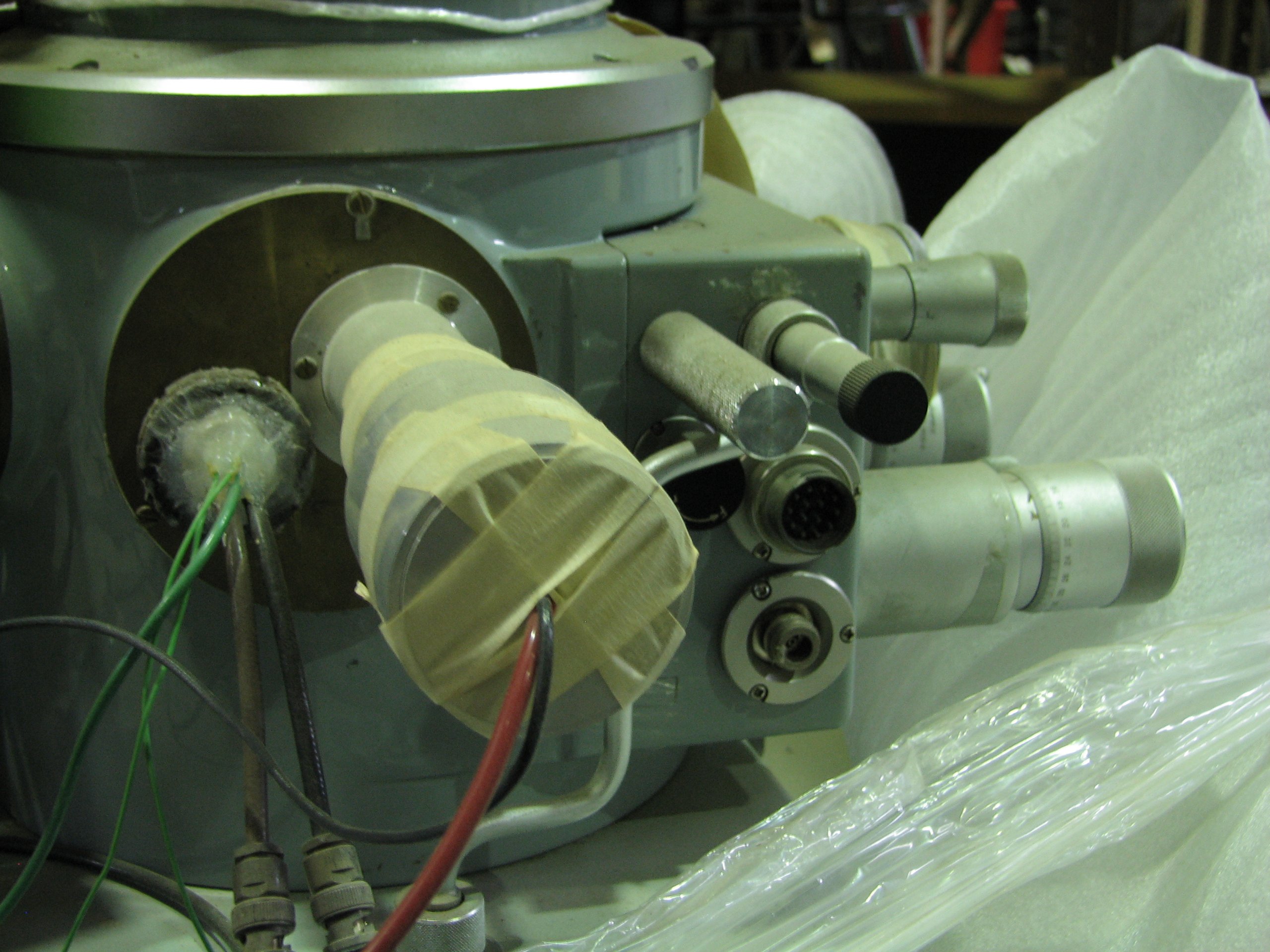
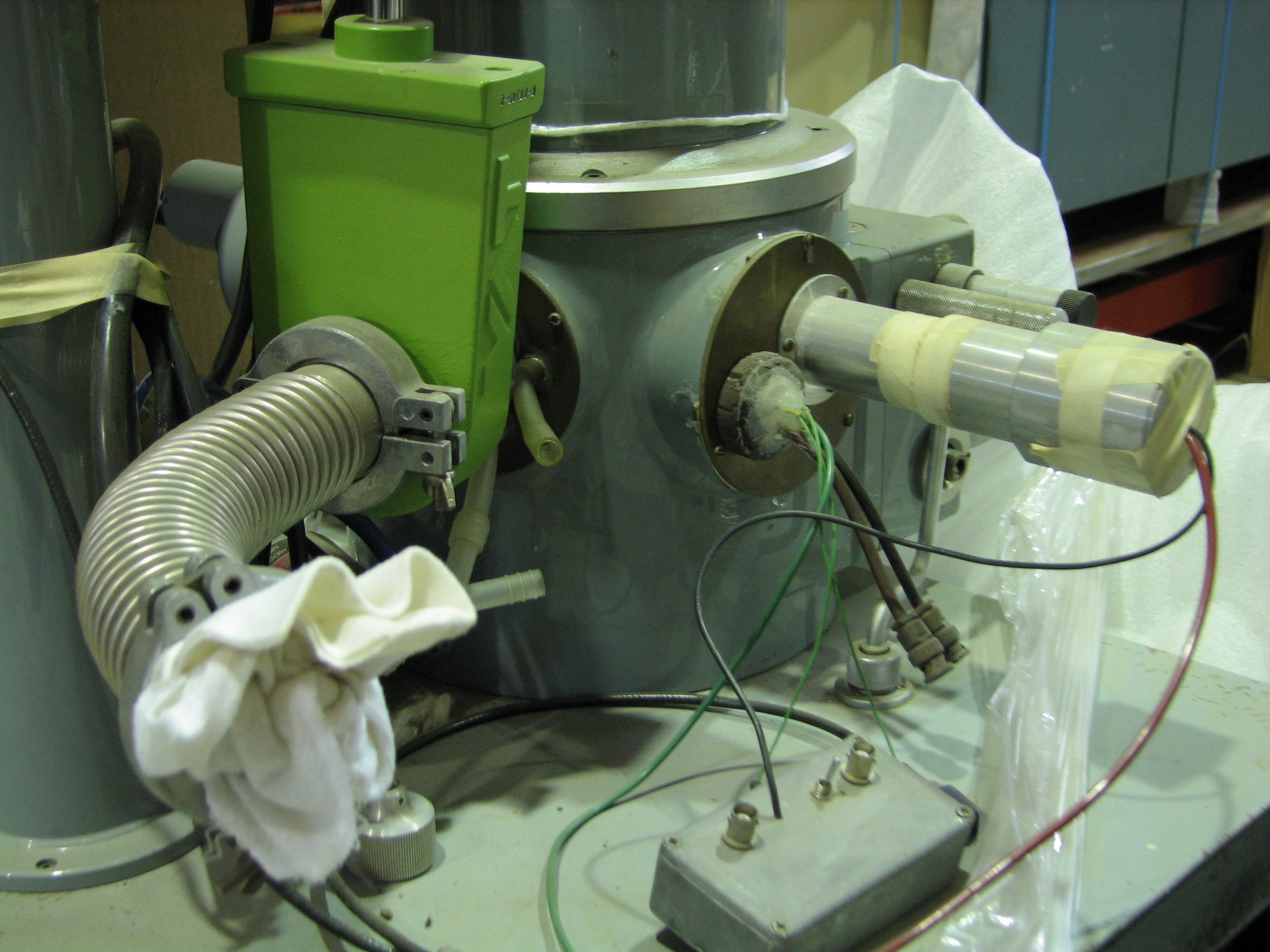
Environmental scanning electron microscope, prototype, steel / aluminium / plastic / rubber/ glass, modified JEOL JSM2 (1968), made by Dr Gerry Danilatos, Australia, 1978 - 2003 The ESEM consists of five main components and numerous accessories. (1) The main console rack houses the electron optical column and specimen chamber. A high-tension electric cable is attached to the top of the electron optical column. The high-tension power supply, in the lower part of the console rack, has inlet and outlet ports for cooling water. An evacuation port attaches the specimen chamber to the two (2 and 3) rotary vacuum pumps. (4) The display and operation system houses the electronic controls for the electron beam, detection devices and imaging systems, such as cathode ray tube viewing screen, and a Polaroid camera to photograph the television image. (5) A power supply box provides stabilised power to the display and operation system. Accessories include metal and plastic specimen holders that allow a specimen to be viewed under environmentally controlled conditions while it is in the path of the electron beam.
PRODUCTION
Notes
This unit, originally a JEOL JSM-2 scanning electron microscope, owned by UNSW, was transferred to CSIRO Wool Research Division, already converted to ESEM mode by Dr. Gerry Danilatos, who then brought it to a state-of-the art prototype just prior to its commercialisation. Commercial prototype models (ElectroScan) based on Danilatos's environmental scanning electron microscope (ESEM) patents derived from this unit, first appeared in 1988. Philips Electronics North America Corporation purchased ElectroScan (11/07/1996) to form Philips Electroscan, which changed to FEI Company (21/02/1997) on merging Philips ElectroScan with FEI. There are now thousands of ESEMs in use world-wide; no research body dealing in biological or environmental mode studies can do without a ESEM.
HISTORY
Notes
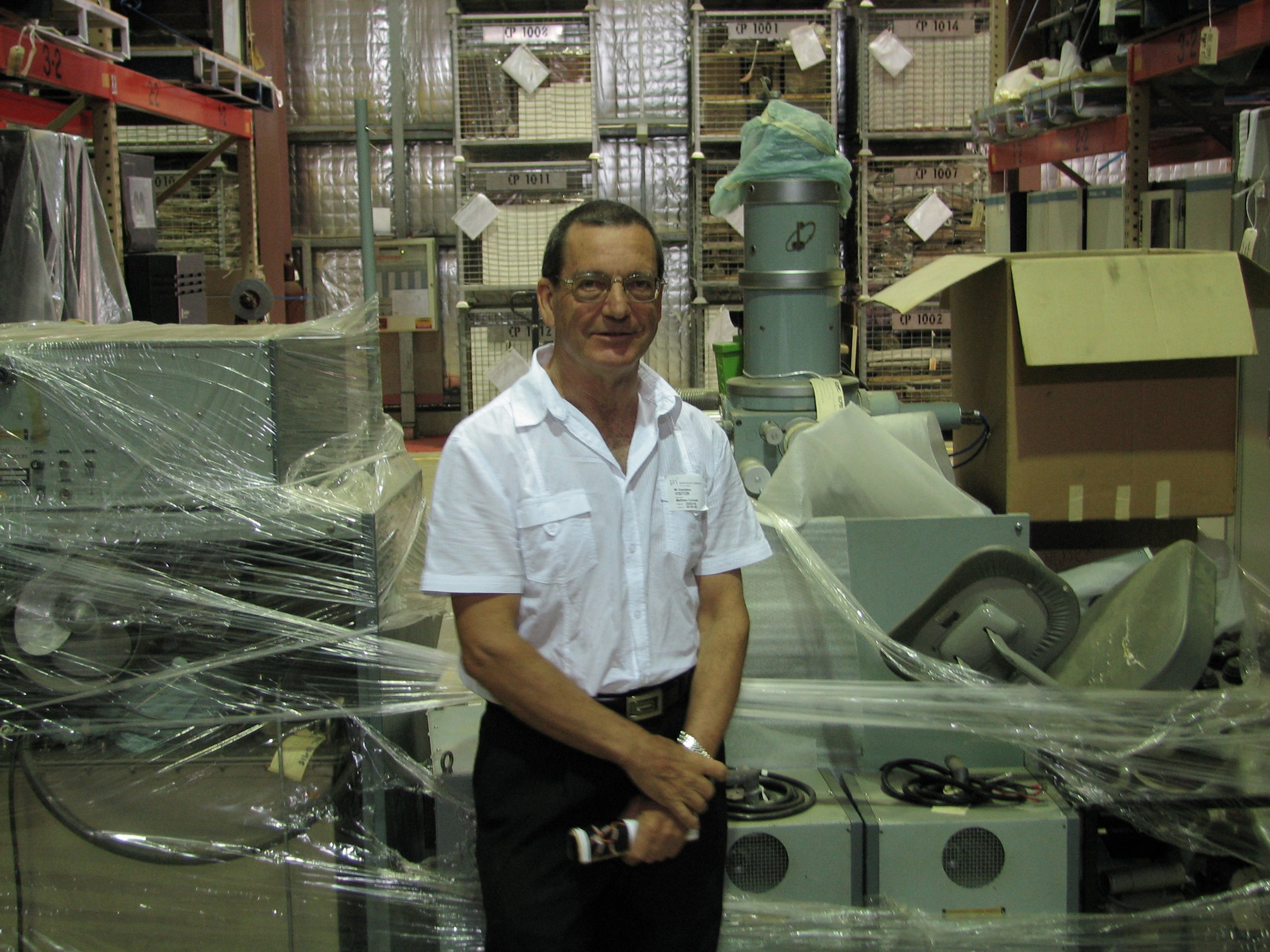
The initial development of the environmental scanning electron microscope (ESEM) took place at the University of New South Wales (UNSW) in the late 1970s; however, support for the program that Danilatos was charting out of his early findings was not adequate. The instrument had to make room for another project and vacate the grounds of the UNSW. With the financial support of the Australian Wool Corporation, the entire activity was transferred to the Textile Physics Division of CSIRO from 1983 to 1986. Even there, ESEM did not develop to its full potential and, once more, Danilatos and his equipment had to leave. At the same time the ElectroScan Corporation had just formed in the USA to manufacture exactly what Danilatos had been working on for all the previous years since 1978. With financial support from the USA, together with all the equipment released by CSIRO to Danilatos on an indefinite loan, the ESEM Research Laboratory was established at North Bondi, NSW, Australia. Due to another relocation the bulky Prototype ESEM could not be maintained beyond 2002 and Danilatos sought to donate it to the Powerhouse Museum for preservation and possible display, after having worked with it for twenty-four solid years.
SOURCE
Credit Line
Gift of Dr Gerry Danilatos, 2010
Acquisition Date
4 May 2010

Copyright for the above image is held by the Powerhouse and may be subject to third-party copyright restrictions. Please submit an Image Licensing Enquiry for information regarding reproduction, copyright and fees. Text is released under Attribution-Non Commercial-No Derivative licence.
Image Licensing Enquiry
Object Enquiry