Papier-mâché anatomical model of a silkworm by Dr Louis Auzoux
Object No. 2795
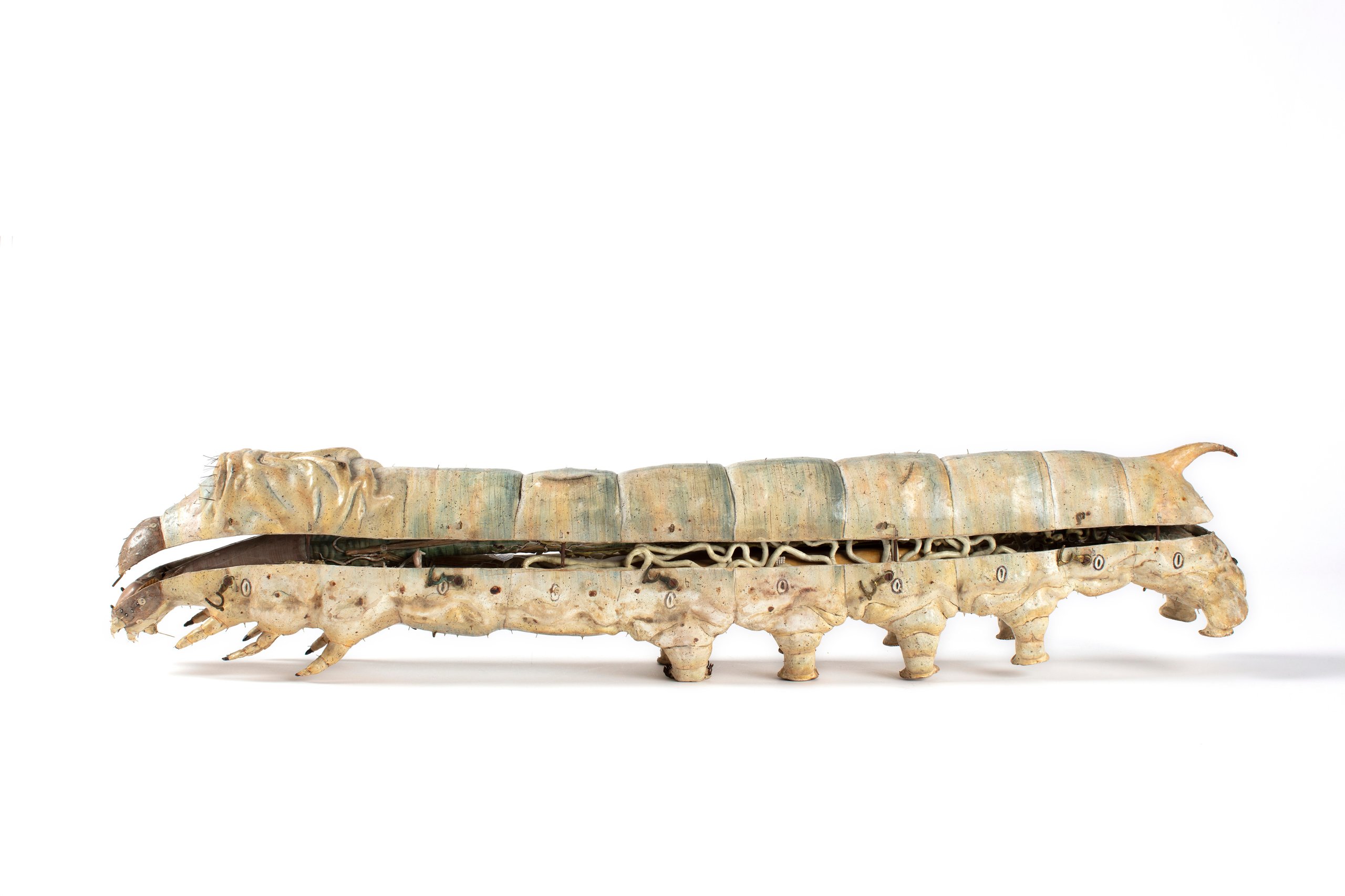
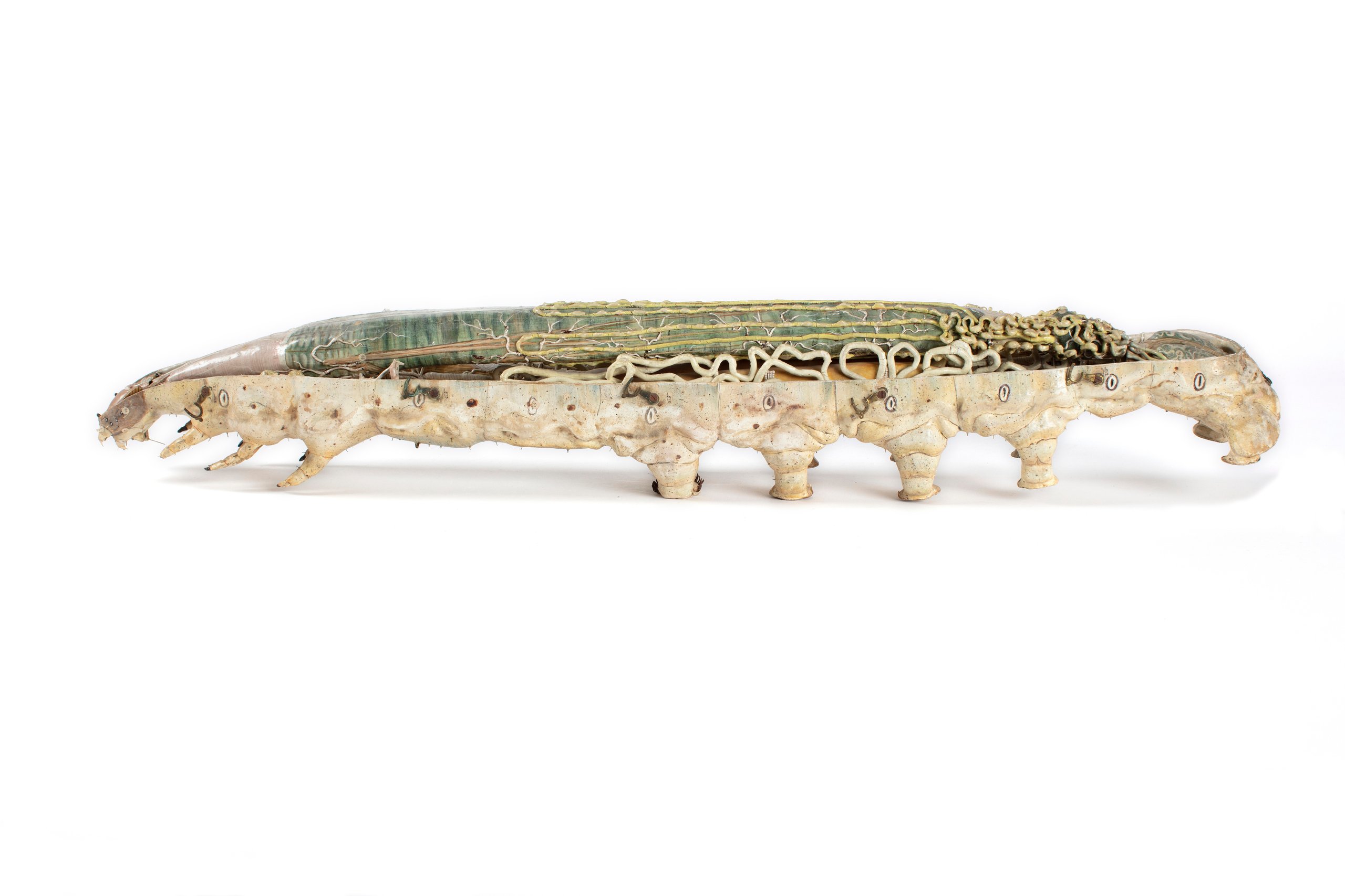
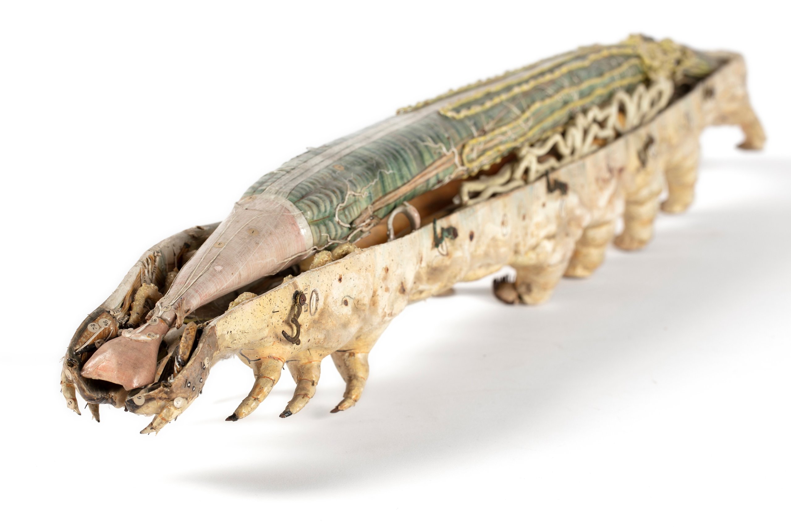
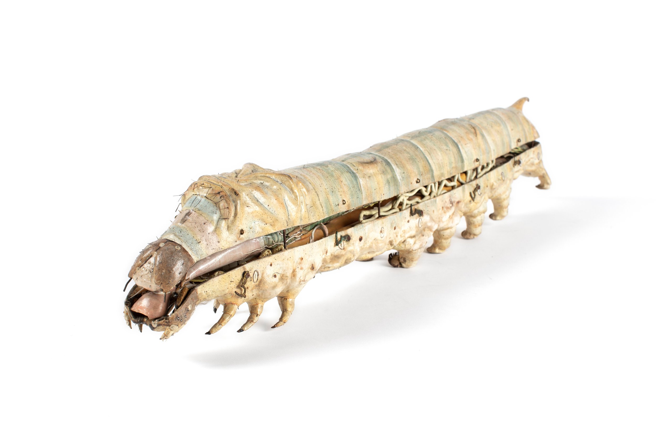
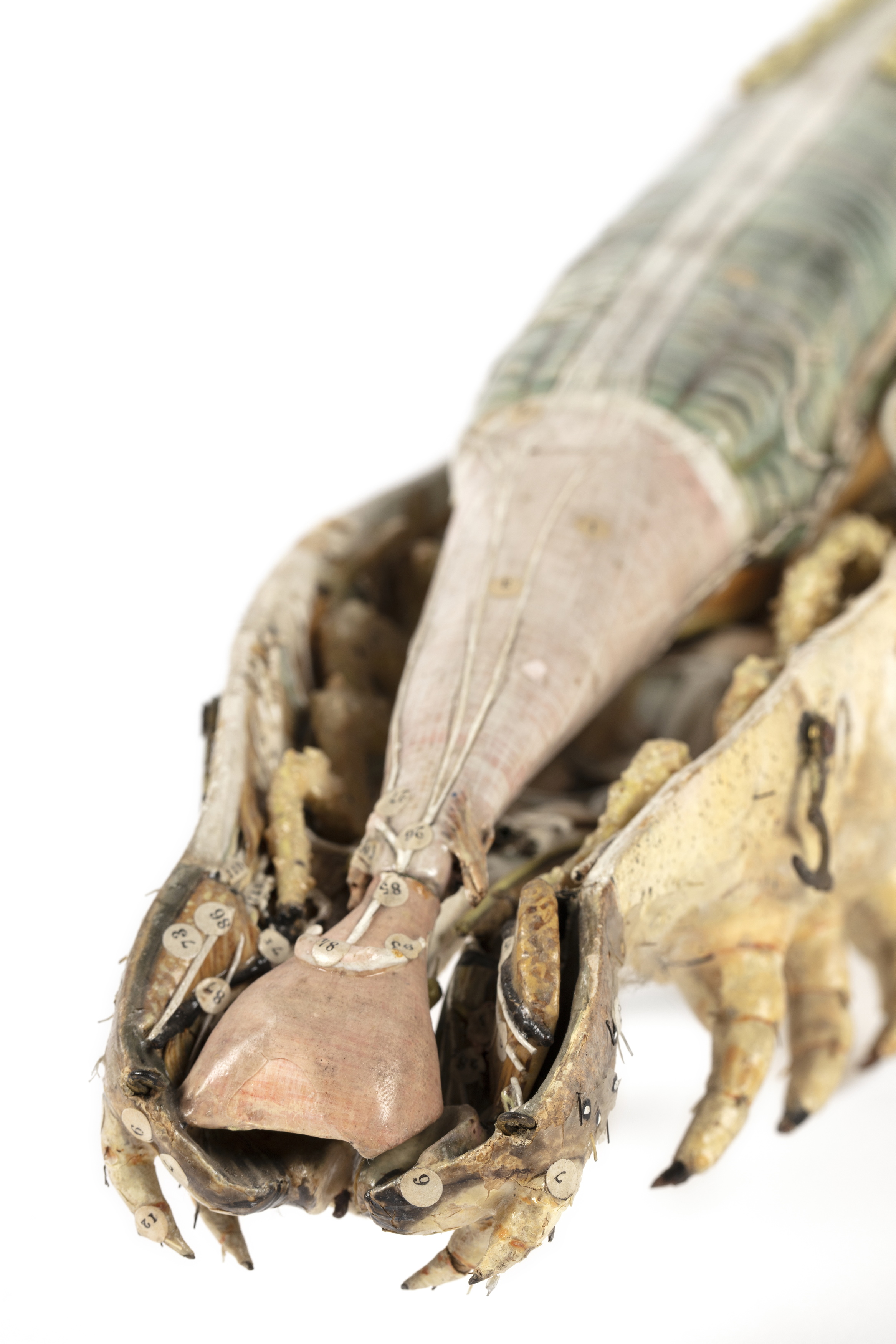
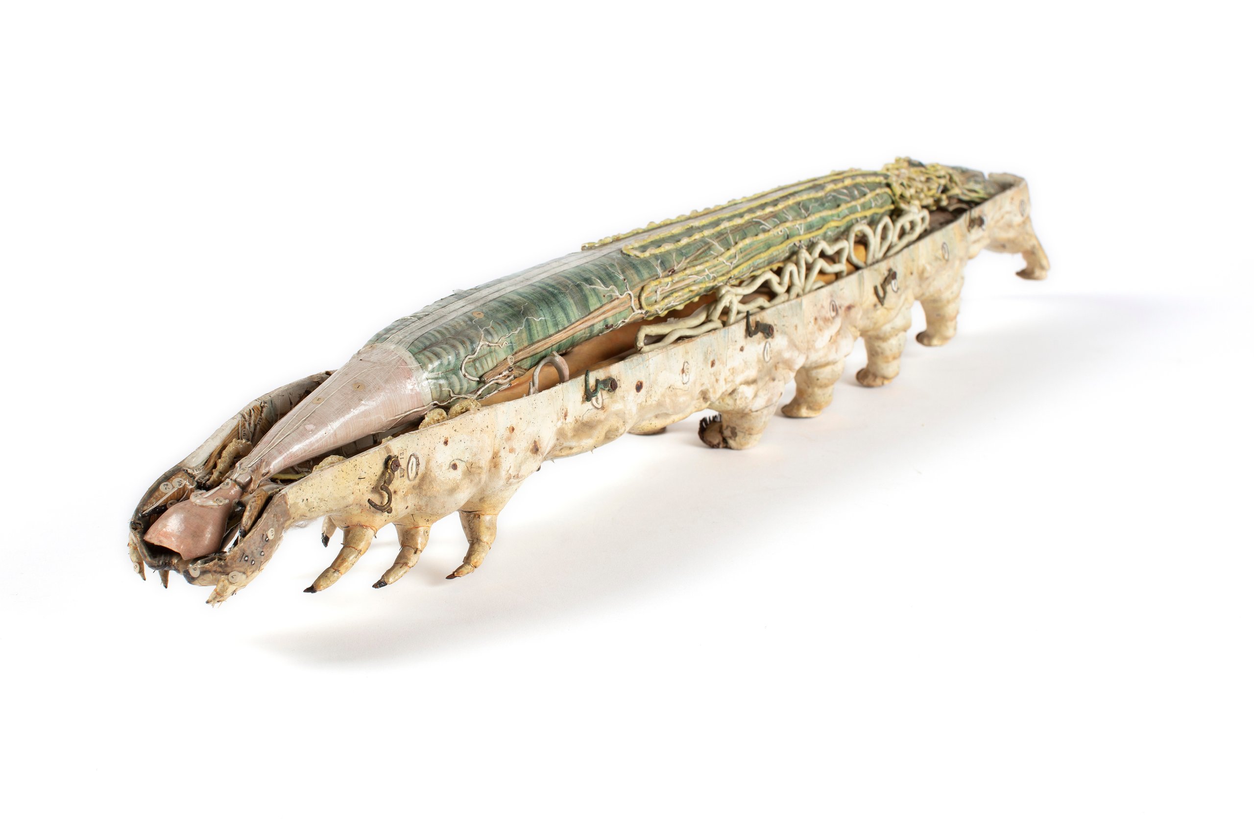
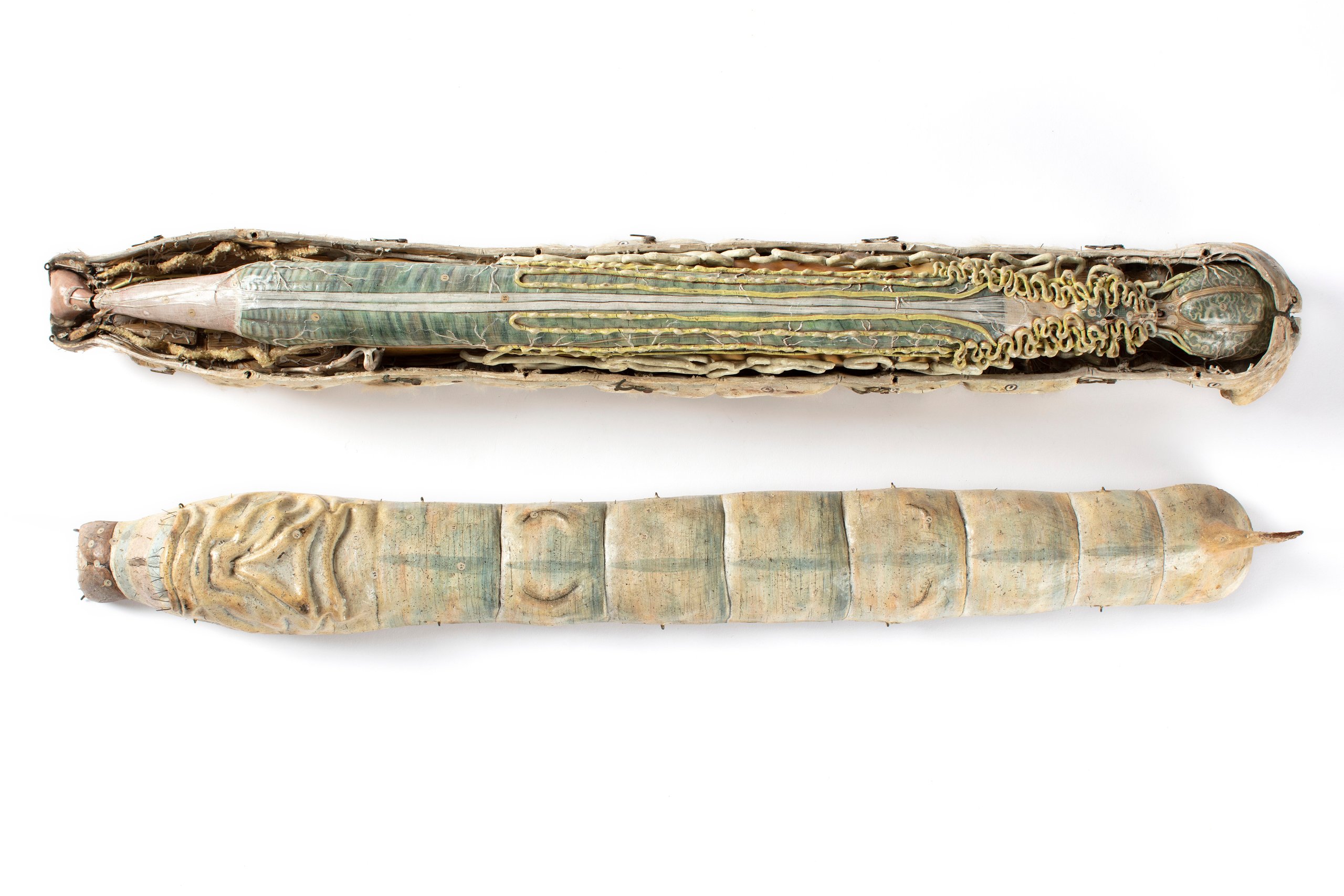
This is an anatomical model of a silkworm made of papier-mache, metal and plaster. It was made by Louis Auzoux in France and purchased by the Museum in 1884. The then curator of the Museum, Joseph Maiden, believed there was great potential for silk cultivation in New South Wales. An extensive display was made of the models which featured different types of silkworms and cocoons, together with production methods and examples of the finished product. Before being displayed in the Museum, this silkworm model 'attracted a great deal of attention' at a Royal Society 'conversazione'. Its five sections can be removed one by one 'to show the internal economy of the worm'. Papier-mache models were introduced in the 1800s as they were more robust than the earlier wax models. It also allowed craftsmen to fashion models in sections which could be removed in layers as if a real dissection were taking place. Louis Thomas Jerome Auzoux was a pioneer of this form of modelling who set up a workshop in his home town of Saint Aubin d'Ecrosville in 1827. His medical background enabled him to make highly accurate models while his experiments with papier-mache resulted in the development of a variety of finishes which incorporated plaster, fabric and glass. The other aspect of Auzoux's success was his application of moulding techniques which allowed him to reproduce his models. A common feature of many of Auzoux's models is the use of paint on a thin plaster layer which covered the papier-mache. Studio artists were employed to add the finishing touches using egg tempura which gave a shiny gloss to the finished work. Iron supports were included to reinforce the delicate areas of some models and metal was sometimes used to connect separate parts. This zoological model of a silk worm was manufactured by the firm run by Dr. Louis Auzoux. It is segmented to allow the removal of the exterior to reveal the organs and tissues inside the silkworm and was made between 1865 and 1884. Its exaggerated size allowed students to easily examine tiny details while the painted colours were often closer to life than the specimens preserved in alcohol which tended to lose their colour. Dr. Auzoux's models were acclaimed throughout Europe and this model was purchased from the German dealer Chrétien Vetter in 1884 some four years after Auzoux had died. Geoff Barker, March, 2007 References Grob, B.W.J., 'The anatomical models of Louis Auzoux', in 'A descriptive catalogue', Colophon, Museum Boerhaave Communication 305, Leiden, Germany, 2004 Scholtz, Gerhard (2005), Better than the real thing? Models - The Third Dimension of Science. Acta Zoologica 86 (4), 303-305, doi: 10.1111/ j.1463-6395.2005.00193.x Chen, Joseph C. T. M.D., Ph.D.; Amar, Arun P. M.D.; Levy, Michael L. M.D.; Apuzzo, Michael L. J. M.D., 'The Development of Anatomic Art and Sciences: The Ceroplastica Anatomic Models of La Specola', Neurosurgery. 45(4):883, October 1999
Loading...
Summary
Object Statement
Anatomical model, Bombyx mori (silkworm), papier-mâché / metal / plaster / paint, made by Louis Thomas Jerome Auzoux, Paris, France, 1833-1884
Physical Description
Anatomical model of a silkworm constructed from papier-mâché and painted plaster with connecting metal hooks. The back of the silkworm can be removed to show the internal organs including the thorax, abdomen and intestines, brain and muscle structure on the underside of the back. On the exterior of the silk worm is shown its mandibles and silk producing glands, three pairs of thoracic legs, four pairs of abdominal prolegs and a pair of anal prolegs. Covering the outside of the body are setae (stiff, strong hairs that help the silkworm to attach itself to surfaces without sliding) and fine particles of silk. The model is painted cream with pale blue, brown, pink and green detail, especially on the internal organs.
DIMENSIONS
Height
150 mm
Width
101 mm
PRODUCTION
Notes
This anatomical model was made by Dr Louis Auzoux no later than 1884, the year this model was purchased. Louis Thomas Jerome Auzoux was born in Normandy in 1797. He obtained a medical degree in 1818 and was appointed to the surgical department of the Hotel-Dieu, with celebrated Dupuytren, the 'Napoleon of surgery.' The shortage of anatomical teaching materials prompted Auzoux, a year later, to begin experimenting with making models. Models in wax were available but were very expensive. In contrast, papier mache was comparatively inexpensive, stable and able to be easily moulded. Furthermore, it was strong enough to allow each model to be taken apart to show the arrangement of organs. Noting the techniques of Parisian doll and puppet makers, Auzoux developed a paper paste, which allowed papier mache models to harden as a solid, supple, light and durable object. This improved upon the early papier mache techniques of Francois Ameline. Auzoux created models, which could be taken to pieces and reassembled, with each part labelled, showing internal anatomy. He called these models 'Anatomie clastique' and designed them for both lay and expert audiences. In 1822 he presented a life-sized model of the human pelvis at the Academie Royale de Medicine, and from 1825 commissions from educational institutions flooded in - requesting human, botanical and veterinary models. Auzoux opened a small factory, in Saint Aubin d'Ecrosville, in 1828, soon employing 100 workers. In 1833, Auzoux established a shop in the rue du Poan in Paris. Over the next century and a half the range increased to some 600 models, the majority zoological and botanical with 100 relating to human anatomy. For many years the Auzoux family had a shop in the Rue du medecine in Paris. The shop finally closed in the 1990s and the contents were sold at auction on 22 October 1998. The models are made with a grey paper pulp, containing granular particles and short fibres. Flax is added to the pulp for models of insect parts, veins and nerves. Auzoux used moulds made from plaster and, later, innovative anatomy moulds for the solid parts of the models. Plaster coats the outside for strength and to provide a base for the paint. The paint is protein-based egg tempera and is protected by a layer of Russian fish glue for models made before 1917, and wood varnish for models made afterwards. The system of labelling was another of Auzoux's innovations: Labels with pointing hands and numbers show where the parts of the model may be disassembled. Anatomical names of the different model parts form a second order in the hierarchy of the anatomy. A third order is the small round numbered labels appearing on some parts, the associated description of which appeared in the accompanying catalogue. Reference: B.W.J. Goob, 'The Anatomical Models of Dr Louis Auzoux' A Descriptive Catalogue (Leiden: Museum Boerhaave Communication 305, 2004)
HISTORY
Notes
Dr Auzoux's factory in Paris produced many different animal models, including insects, amphibians, reptiles, birds, wild and domestic mammals, and botanical specimens. His models were popular for use in teaching and museums developed exhibits comparing the differences between botanical models, human and veterinary structures.
SOURCE
Credit Line
Purchased 1883
Acquisition Date
23 November 1883

Copyright for the above image is held by the Powerhouse and may be subject to third-party copyright restrictions. Please submit an Image Licensing Enquiry for information regarding reproduction, copyright and fees. Text is released under Attribution-Non Commercial-No Derivative licence.
Image Licensing Enquiry
Object Enquiry